The picture on the left shows every detail in a fully assembled catheter connection in the optical tomogram.
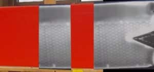
The image in the middle shows a thermal tomogram. Shown is the honeycomb construction under the planking inside an aircraft wing.
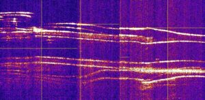
The image on the right shows a radar tomography ridge. The radar tomogram shows the individual layers of built-in reinforcing iron in a ceiling and at the same time built-in electrical cables or pipes for floor heating.
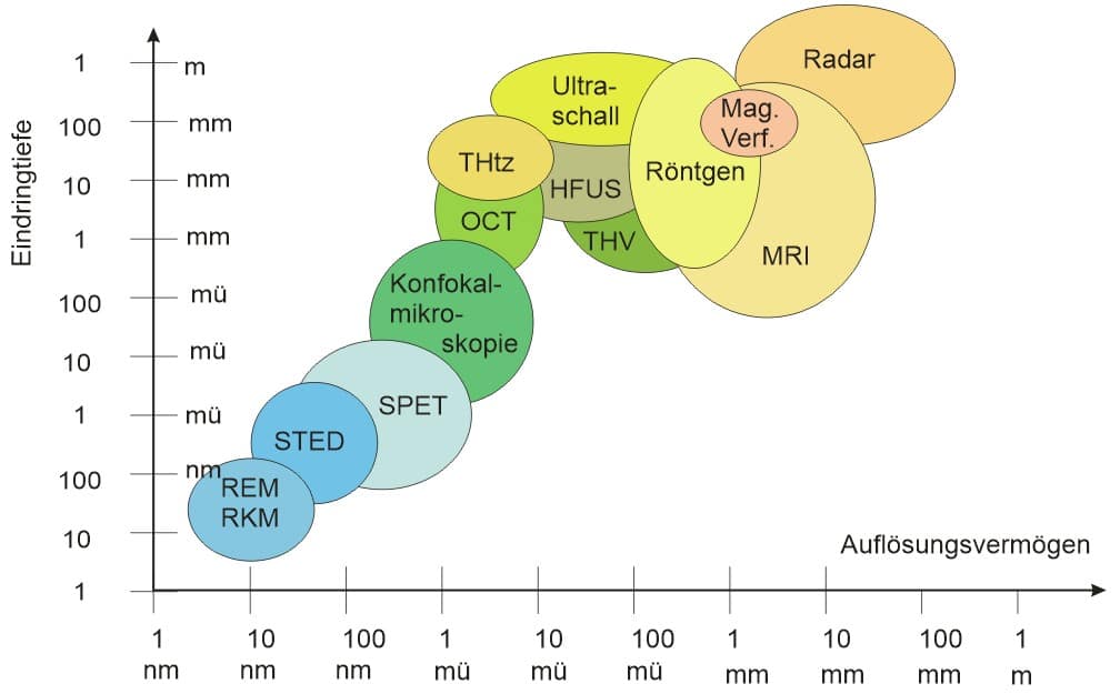
Radar: Different frequency bands
Mag.Verf.: Magnetic methods
MRI: magnetic resonance procedure
THV: Thermography
HFUS: High Frequency Ultrasound
THtz: Therahertz method
OCT: Optical Coherence Tomography
SPET: Stimulated Photon Emission Tomography
STED: Stimulated Phote Thinning
REE, RKM: scanning electron microscopy, atomic force microscopy
irscat.ch GmbH
Gottfried Kellerstrasse 2
6010 Kriens
Switzerland